
Article
Methylene blue and brain-support cells: astrocytes, microglia, and inflammation
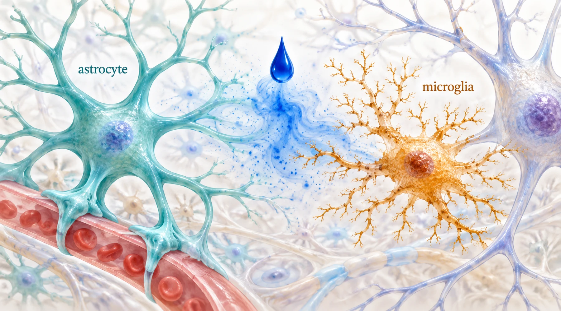
Suppose you run two experiments with the same blue dye and get two encouraging results. In one dish, star-shaped support cells survive oxygen and glucose loss. In another, immune cells of the brain keep their calm branched shape despite a bacterial toxin. It is tempting to combine the two into one claim: the dye calms brain inflammation and protects the brain. That combination is exactly where careful readers should slow down.
The brain has two support populations with different jobs, and methylene blue meets each of them through a different experimental trigger. Astrocytes are metabolic support cells. They sit between blood vessels and neurons, clear excess glutamate, buffer potassium, store glycogen, and shuttle fuel to neurons when demand spikes. Microglia are the resident immune surveyors. Their fine processes patrol tissue, detect bacterial and damage signals, and on alarm retract into a rounded amoeboid form built for migration and engulfment while releasing signaling molecules. A result in one population does not transfer to the other, and the measurement used in each case decides what was actually shown.
Astrocytes under metabolic stress: timing is the result
The astrocyte experiment starts with deprivation. Researchers cultured cortical astrocytes from newborn mice, removed glucose and nearly all oxygen for six hours, then restored both for twenty four hours, a laboratory model of stroke followed by reperfusion. When methylene blue was present during the deprivation phase, cell survival got worse. When it was added only during the reoxygenation phase, at 0.1 and 1 micromolar, survival improved significantly by a calcein viability assay. The mouse astrocyte study reports this timing split alongside the respiration data.
That split is the finding that matters most. During deprivation there is almost no oxygen to accept electrons, so an electron relay has nowhere to deliver. During reoxygenation the machinery restarts and the dye has work to do. Treated astrocytes consumed more oxygen, took up more glucose, produced more ATP, and rebuilt glycogen stores, with higher hexokinase activity and no rise in mitochondrial superoxide. The proposed explanation is the familiar one from the account of methylene blue as a mitochondrial electron carrier, shuttling electrons past blocked points in the respiratory chain so respiration restarts cleanly rather than leaking electrons into reactive species.
Note what kind of evidence this is. It is a survival and metabolism result in one isolated cell type, with a named effective window (recovery, not deprivation) and a named concentration range. It says nothing about immune signaling, and nothing about behavior.
Microglia under bacterial alarm: metabolism reprogrammed
The microglia experiments start from the opposite trigger, not starvation but a false infection. Lipopolysaccharide, a fragment of bacterial outer membrane, binds innate immune sensors and convinces microglia that pathogens have arrived, even though no live bacteria are present. In mice given lipopolysaccharide and in a cultured microglial cell line, the result is a familiar inflammatory program: pro-inflammatory factors rise, microglia polarize toward the aggressive M1 state, neighboring neurons die, and the animals perform worse on learning and memory tasks.
The murine study of this response traces it to an oxygen-sensing pathway: lipopolysaccharide drives overexpression of PHD3, one of three prolyl hydroxylase enzymes, through an upstream Siah2/Morg1 chain. At the same time the cells reprogram their energy use toward aerobic glycolysis with elevated mitochondrial reactive oxygen species, a shift the authors measured directly with Seahorse metabolic assays of oxygen consumption and extracellular acidification. Methylene blue reversed both the metabolic shift and the PHD3 overexpression, lowered the inflammatory factors, reduced neuronal death, and improved the behavioral scores. Positron emission tomography added an imaging layer: lipopolysaccharide raised hippocampal glucose uptake on FDG-PET, and treatment brought it back down, consistent with a calmer inflammatory metabolism in the living brain.
This is a richer measurement set than the astrocyte study (cytokines, metabolism, imaging, behavior in animals), but it belongs to a different trigger and a different cell. Readers comparing the two should keep the triggers apart: one study rescues starving support cells during recovery, the other damps a false infection alarm in immune cells.
Microglia under the microscope: shape as a readout
A third experiment looks at the same bacterial alarm through shape alone. In glial cultures from rat cortex, control microglia stained for the marker IBA1 show many fine processes, the branched surveying form. After twenty four hours of lipopolysaccharide at 10 micrograms per milliliter, the cells flatten into the amoeboid form, and their cell body area grows while the perimeter stays roughly constant, which is the geometric signature of rounding up. Methylene blue at 1 micromolar, given alongside the toxin, preserved the branched processes and kept body area and perimeter near control values. The rat culture study quantifies this shape rescue with cell body area and perimeter measurements.
Two details make this study worth more than a simple picture. First, the comparison compound menadione, another redox-active molecule at the same concentration, did not rescue shape and by itself pushed cells toward the activated form, so the effect is not a generic property of all redox cyclers. Second, the authors link the dye's effect to Nrf2 activation, a transcription factor program behind antioxidant and anti-inflammatory genes, rather than to direct scavenging alone. Related coverage of redox and inflammatory signaling puts this Nrf2 connection in context.
Shape is nevertheless the thinnest of the three readouts. A branched cell is usually a surveying cell and a rounded cell is usually an activated one, but morphology is a proxy, not a transcript. This culture contained no blood-borne immune cells, no circuitry, and no behavioral output.
Reading the three results side by side
| Trigger | Cell type | What was measured | What methylene blue did |
|---|---|---|---|
| Oxygen and glucose removed, then restored | Mouse astrocytes | Viability, oxygen consumption, glucose uptake, ATP, glycogen, superoxide | Protected only when given during reoxygenation; raised respiration and stores without raising superoxide |
| Bacterial toxin fragment | Mouse microglia, in culture and in live mice | Cytokines, M1 markers, Seahorse metabolism, PHD3 pathway proteins, FDG-PET, memory tasks | Reversed glycolytic reprogramming and PHD3 overexpression; lowered inflammation; improved behavior |
| Bacterial toxin fragment | Rat microglia in mixed glial culture | IBA1 body area, perimeter, process branching | Preserved branched resting shape; menadione did not |
The pattern across rows is that each trigger gets its own measurement and its own answer. Metabolic rescue answers a metabolism question. Cytokine and behavior data answer an inflammation question. Shape answers a shape question. None of the three crosses over on its own.
What calmer-looking microglia do not prove
This is the sentence the whole topic needs: less activated-looking microglia, or altered microglial metabolism, does not by itself prove improved brain function.
Morphology can change without function following. A cell can keep its branches while still releasing cytokines, or round up while doing useful debris clearance. Metabolic readings have the same limit: a Seahorse trace or a PET scan shows energy handling, not cognition. The mouse study that paired metabolic and shape-like readouts with behavioral tests is stronger precisely because it stacked levels of evidence instead of asking one measurement to carry the conclusion. Even there, the usual limits apply, since lipopolysaccharide models a sterile alarm rather than a real infection, mice are not people, and the authors flag species differences themselves. Anyone planning follow-up work should weigh how microglial findings translate across models before assuming the pathway behaves identically in human tissue.
A practical corollary follows for timing. The astrocyte result punishes early dosing and rewards dosing during recovery. The microglia results come from dosing around the inflammatory trigger. Brain injury in real life mixes both phases at once: energy failure plus immune alarm, evolving over hours. An experiment that isolates one phase cannot set the schedule for the combined case, which is why discussions of dosing windows keep the deprivation and inflammation timelines separate.
The honest summary fits in one paragraph. In isolated astrocytes, methylene blue given during reoxygenation restarts respiration and saves cells. In microglia challenged with a bacterial fragment, it reverses an inflammatory metabolic program through a specific PHD3 pathway and preserves resting shape. Each result is real within its setup, and neither is a general verdict on brain inflammation. The glial comparison is useful exactly because it stops the two stories from blurring into one.