
Article
Why methylene blue is used as an acid-fast counterstain
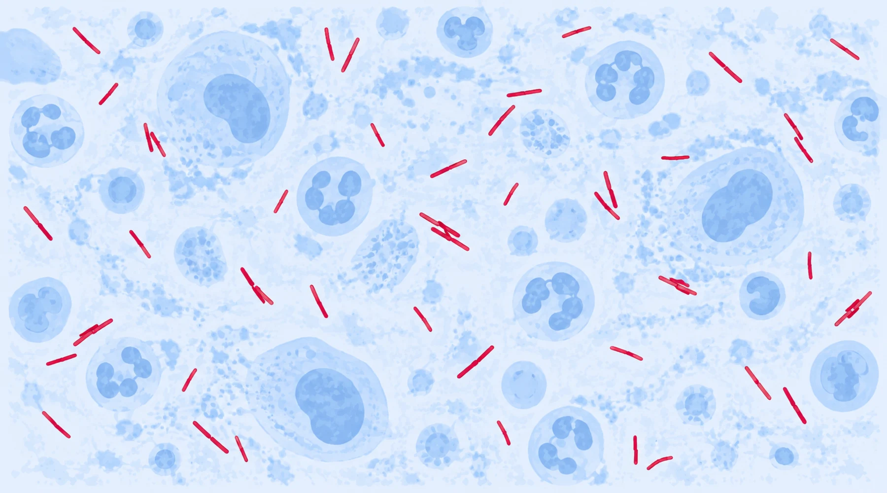
A technician examines a sputum smear after the full staining sequence. Red rods stand out against a blue field, and the report goes out as acid-fast bacilli seen. The next day a different slide shows a uniform blue field with no red rods, and it gets reported as negative for tuberculosis. One of these reports claims more than the slide supports, and it is not the first one.
The confusion is conceptual. The technician treated the blue dye as part of the detection chemistry, as if methylene blue helped find the mycobacteria. It did not. By the time methylene blue touches the slide, the test is already over. The dye only paints the background so the result is readable. This article explains the purpose of methylene blue in acid-fast staining, why it is blue rather than any other color, and what red against blue licenses you to conclude.
The short answer
Acid-fast staining is a differential test built from three ideas. Carbol fuchsin, the primary stain, is forced into every cell including waxy mycobacteria. Acid-alcohol, the decolorizer, strips the red dye out of ordinary cells but fails to extract it from acid-fast ones. Methylene blue, the counterstain, then colors everything the decolorizer emptied, so non-acid-fast cells, host cells, and debris appear blue while acid-fast bacilli stay red. The counterstain supplies contrast. It plays no part in the chemistry that makes a cell acid-fast.
What each step does
The diagram below shows the division of labor. Read it left to right as a procedure, and read the bottom strip as a reminder that each staining family asks a different question.
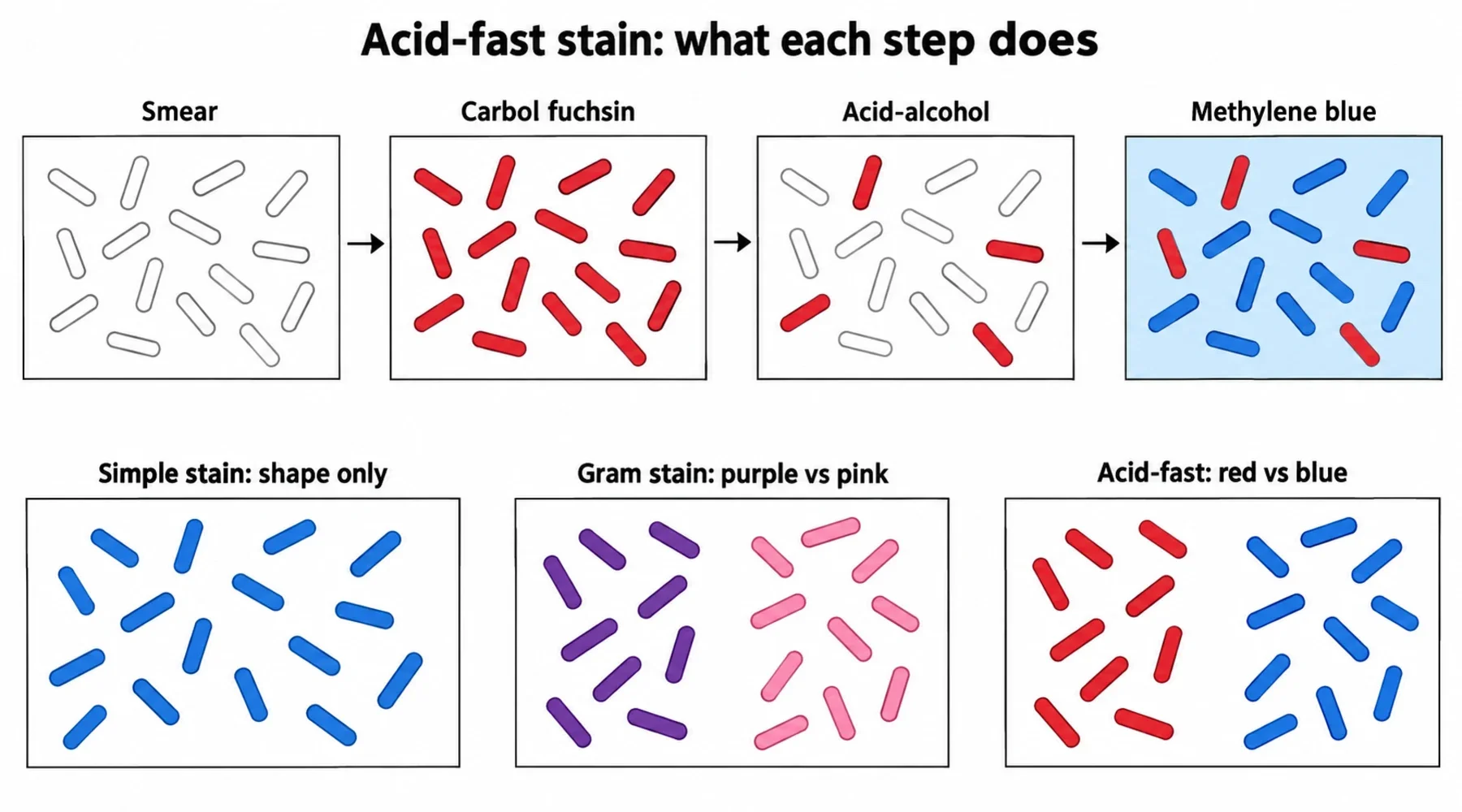
Penetration comes first. Mycobacteria resist ordinary aqueous dyes because their envelope is unusually hydrophobic, so the protocol forces entry. In the Ziehl-Neelsen method, heat steaming drives phenol-containing carbol fuchsin through the waxy wall. In the Kinyoun cold method, heat is replaced by a far stronger primary stain formulation, and some modified heatless versions add surfactants. The American Society for Microbiology acid-fast stain protocols document both formulations side by side, and the concentration difference is striking: roughly 0.3 g basic fuchsin per 100 mL for the heated method versus about 4 g for the cold method.
Differentiation comes second. Acid-alcohol, commonly 3 mL concentrated hydrochloric acid in 97 mL of 95 percent ethanol, washes the red dye out of every cell whose wall offers no special resistance. Cells with the intact mycobacterial lipid envelope hold onto the dye. This is the step the whole method turns on, and the step most sensitive to technique, as discussed below.
Contrast comes last. The slide now holds red acid-fast cells on a nearly colorless background of emptied cells. Methylene blue at 0.3 percent w/v restores visibility to that background in a color maximally distant from red. Counterstaining is brief, typically 20 to 30 seconds in the heated method and up to a few minutes in some cold-method variants, and timing is protocol dependent rather than a property of the dye itself.
Why this dye, and why blue
Methylene blue is a cationic basic dye, which means its colored ion carries a positive charge and binds the negatively charged surfaces of decolorized bacteria, host cells, and debris. OpenStax Microbiology describes this charge logic as the general principle behind basic stains. But methylene blue is not chemically unique here. Crystal violet and safranin are basic dyes too. The choice is optical, not chemical.
Red rods need a background in a contrasting hue, and blue sits opposite red in the most useful way for bright-field microscopy. Safranin would be a poor counterstain because its pink-red endpoint sits too close to retained carbol fuchsin, collapsing the very distinction the procedure created. Crystal violet would give a dark violet background that competes with thin red bacilli for attention. The strongest evidence that the job is contrast rather than chemistry is substitution: green counterstains such as malachite green and brilliant green serve in some veterinary and modified protocols and the test still works, because any clearly different hue does the same job.
Two practical corollaries follow. First, the standard recipe specifies no pH adjustment; do not confuse this plain aqueous 0.3 percent solution with alkaline formulations such as Loeffler's methylene blue, which belong to a different staining concept. Second, a heavy hand hurts. Histology literature specifically warns that an overly dense blue background can mask scanty bacilli, so flooding the slide longer than the protocol allows trades readability for nothing.
The wall chemistry that makes the test possible
The reason acid-alcohol discriminates lies in the mycobacterial envelope. Its structural core is the mycolyl-arabinogalactan-peptidoglycan complex: peptidoglycan linked to arabinogalactan whose outer ends carry very long chain mycolic acids, forming a lipid rich outer mycomembrane of unusually low permeability. The review of the mycobacterial outer membrane details this architecture. The barrier works in both directions. It resists dye entry, which is why penetration needs heat or a strengthened primary stain, and it resists dye extraction, which is why acid-alcohol fails to remove carbol fuchsin once it is inside. Ordinary bacteria lack this envelope and lose the dye. Acid-fastness is therefore best understood as a lipid barrier phenomenon, not as fuchsin chemically binding to mycolic acid.
This chemistry also explains a common puzzle: mycobacteria stain poorly and unpredictably by the Gram method, sometimes appearing as faint beaded forms or unstained ghosts. Their hydrophobic wall limits crystal violet penetration. Acid-fast staining is not a modified Gram stain. It is a different differential principle built around a different wall property, as the simple stain versus Gram stain comparison shows for the other two methods.
Three methods, three questions
| Method | Test performed | Positive endpoint | What the result proves |
|---|---|---|---|
| Simple stain (e.g. methylene blue alone) | None; one dye colors all cells | Blue cells of a given shape | Morphology only: shape and arrangement, no classification |
| Gram stain | Alcohol or acetone challenge of the crystal violet iodine complex | Purple (positive) vs pink (negative) | Envelope behavior under solvent extraction; a classification into two wall types |
| Acid-fast stain | Acid-alcohol challenge of carbol fuchsin forced into waxy walls | Red (acid-fast) vs blue (non-acid-fast) | Retention against acid-alcohol; acid-fastness, which is consistent with mycobacteria but is not species identification |
For the wider family of one-dye bacterial methods, see the microscopy staining atlas.
What red against blue cannot prove
Red rods mean the organisms retained carbol fuchsin through acid-alcohol. That finding is consistent with Mycobacterium tuberculosis, but CDC tuberculosis diagnostic guidance notes explicitly that other mycobacteria are also acid-fast, so species identification needs culture and nucleic acid testing, not color.
Technique errors cluster around the two washes. Under-decolorization leaves background material red and invites false positive reads. Over-decolorization strips weakly retained primary stain and contributes to false negatives. A counterstain applied too heavily buries scanty bacilli in blue. Thick smears, precipitated reagents, and contaminated rinse water add their own artifacts.
Sensitivity is bounded by organism burden no matter how clean the technique. Conventional smear microscopy needs roughly 5,000 to 10,000 bacilli per milliliter of sputum, and older CDC evidence reviews placed smear sensitivity around 45 to 80 percent among culture confirmed pulmonary cases. A negative smear therefore never excludes tuberculosis. Current CDC guidance keeps culture alongside every smear and recommends nucleic acid amplification testing on at least one respiratory specimen when pulmonary tuberculosis is considered and the result would change management.
Practical work belongs to authorized protocols
Teaching descriptions, including this article, do not authorize clinical work. For bench practice, follow the laboratory's validated standard operating procedure aligned with current CDC, APHL, or CLSI documents; the CDC acid-fast staining reference held in its Stacks archive shows how such procedures specify reagents and readouts. Tuberculosis specimen work requires containment: smears are prepared in a biological safety cabinet because preparation generates aerosols and splatter, and CDC laboratory safety guidance states that even heat fixed smears may contain viable tubercle bacilli and must still be handled as contaminated. The CDC Public Health Image Library entry for a carbol-fuchsin and methylene blue smear examined for M. leprae is a useful reminder of the method's range beyond tuberculosis: the same red on blue contrast serves wherever acid-fast organisms are sought, and a careful negative read is itself a result worth recording.
Reading the finished slide
The counterstain earns its place during screening. A microscopist scanning at low power first uses the blue background to judge smear quality: even cell distribution, intact host cells for focus, no heavy precipitate. Red candidates then draw the eye precisely because nothing else on the slide is red. Confirmation happens under oil immersion, where true bacilli show the expected thin, sometimes slightly curved or beaded rods rather than uniform dye debris.
Background staining also sets the vocabulary of the report. A slide with red bacilli is reported as acid-fast bacilli seen, often with a semiquantitative grade per the laboratory's scale. A slide with no red organisms is reported as no acid-fast bacilli seen, never as negative for tuberculosis. The distinction matters because the blue field proves the stain worked, cells took up counterstain and optics were adequate, while saying nothing about organisms below the detection limit. A blank blue field is a valid result about the slide, not a verdict about the patient.
Remember the division of labor and the reports write themselves. Carbol fuchsin detects candidates. Acid-alcohol runs the test. Methylene blue only makes the answer legible: red kept the dye against acid, blue lost it. Everything else, species, burden, and clinical meaning, comes from work beyond the slide.