
Article
Loeffler's alkaline and polychrome methylene blue stains explained
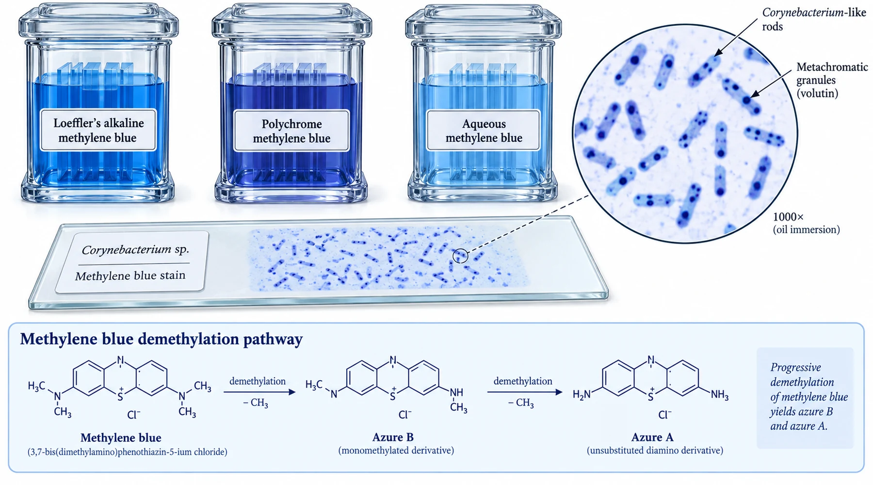
Three bottles can sit side by side on a staining bench, each labeled with some variant of methylene blue. One says aqueous methylene blue 1%. One says Loeffler's alkaline methylene blue. One says polychrome methylene blue. A busy worker reaches for whichever is closest. The smear then stains too faintly, or too uniformly blue, or in unexpected shades of purple, and the result gets blamed on the sample.
This is a formulation problem, not a sample problem. The three solutions contain related dyes but embody different staining concepts. One is a plain cationic dye at near neutral pH. One is the same dye made more aggressive by alkali. One is no longer a single dye at all, but a deliberately oxidized mixture. Use the wrong one and the staining principle changes under your hands.
The short answer
Plain aqueous methylene blue is the baseline: about 1% dye in water, a simple cationic stain for bacterial morphology and general microscopy. Loeffler's alkaline methylene blue adds a small measured amount of dilute alkali to an alcoholic stock diluted in water, which raises the pH and drives the cationic dye harder onto acidic bacterial structures. Polychrome methylene blue is methylene blue that has been deliberately ripened by oxidation in alkali, so the bottle contains a mixture of demethylated homologues (azure A, azure B, azure C), thionin, and methylene violet alongside residual methylene blue, and that mixture stains different structures in different hues.
If you remember only one sentence: alkalinization changes staining intensity, while polychroming changes staining colors.
Ordinary aqueous methylene blue: the reference point
The simplest formulation is methylene blue dissolved in water, commonly at 1% w/v, without alkali and without oxidation. The colored species is the methylthioninium cation with a chloride counterion, a small flat phenothiazine dye described in PubChem's methylene blue record as methylthioninium chloride. As a cation it binds phosphate backbones of nucleic acids and acidic groups on bacterial cell walls, coloring cells blue against a light background.
Its virtue is predictability. Nuclei and bacteria appear blue. Contrast is modest. Faint organisms may stain weakly, which is exactly the limitation Loeffler set out to fix. For the broader family of simple bacterial stains this solution belongs to, see an overview of bacterial simple staining. Identity discipline still matters here: methyl blue is a different molecule entirely, as methyl blue versus methylene blue explains, so a catalog search on color name alone can deliver the wrong dye class.
Loeffler's alkaline methylene blue: the same dye, pushed harder
The classic Loeffler recipe combines an alcoholic methylene blue stock with dilute alkali in water. Textbook versions call for 0.3 g of dye in 30 mL of 95% ethanol plus 100 mL of 0.01% aqueous potassium hydroxide (or equivalently 30 mL of saturated alcoholic methylene blue, 100 mL of distilled water, and 1 mL of 1% KOH), giving a working solution near 0.3% dye at distinctly alkaline pH. Commercial versions follow the same principle, and the Thermo Fisher microbiology stains catalog lists Loeffler's alkaline methylene blue alongside polychrome and aqueous variants as separate catalog items with separate intended uses.
The alkali is the active idea. Raising the pH deprotonates carboxyl and phosphate groups on the specimen, increasing the density of negative sites, while the ethanol stock keeps the dye fully dissolved so it arrives at those sites in monomeric form. The result is faster, more intense staining of bacteria than neutral aqueous dye gives, with the familiar deep blue rods and cocci of a routine bacteriological smear. Staining time is typically one to three minutes, followed by a water rinse.
The classic application is Corynebacterium diphtheriae grown on Loeffler serum medium: slender, often clubbed rods with strongly stained metachromatic Babes-Ernst granules of stored polyphosphate. The pairing of medium and stain is historical, both credited to the same bacteriologist, and the granule contrast is the diagnostic payoff. Beyond diphtheria work the stain serves as a general bacteriological stain for smears from cultures, for Neisseria morphology in clinical smears, and wherever a Gram stain is unnecessary but plain aqueous blue proves too faint. It is a monochrome stain. Everything acidic goes some shade of blue. When a protocol calls for differential color, alkali alone cannot supply it.
One practical caution: alkaline dye solutions deteriorate and precipitate with storage and carbon dioxide uptake. Laboratories either prepare modest volumes or buy the catalog solution and respect its shelf life rather than nursing an old bottle that now throws sediment onto every slide.
Polychrome methylene blue: one name, several dyes
Polychrome methylene blue starts from the same dye but subjects it to controlled oxidation in alkali, historically called ripening or polychroming. Alkaline aging, heating with carbonate, or oxidation with agents such as dichromate strips methyl groups from the dimethylamino substituents stepwise. The product is a mixture: residual methylene blue plus azure B (one methyl lost), azure A and azure C (further demethylation), thionin (fully demethylated), and oxidative side products of the methylene violet family. Each component has its own hue and tissue affinity, so the mixture stains metachromatically: nuclei, granules, mucins, and cartilage each take distinct blue to red-purple tones.
This chemistry is the same chemistry behind the famous blood stains. Early workers found that alkalinized methylene blue stained malaria parasite nuclei in shades of purple, but their aqueous ripened solutions were unstable and precipitated within days. The review of how the purple blood stain was invented traces how Malachowski and Romanowsky independently exploited this effect in 1891, how methanol was introduced to stabilize the dye precipitate and fix films without heat, and how Giemsa later standardized the mixture around measured azure B plus eosin with glycerol stabilization. Modern Giemsa-type stains descend directly from polychrome methylene blue plus eosin, the combination that produces the Romanowsky effect. For the eosin pairing and the azure B component in detail, see Romanowsky-type stains and the azure B eosin interaction.
Because it is a mixture, polychrome methylene blue is the least standardized of the three by nature. Its exact hue balance depends on the oxidation route and extent, which is why histology suppliers sell it as a distinct product with its own performance testing rather than as aged Loeffler's. Typical uses include metachromatic demonstration of mast cell granules, mucins, and cartilage, plasma cell detail, and Helicobacter pylori in tissue sections by Goodpasture-type polychrome methods. It is the right choice when the question is differentiation by color, and the wrong choice when the protocol expects a clean monochrome bacterial silhouette.
Formulation and purpose comparison
| Formulation | What is in the bottle | pH character | Staining principle | Use it when |
|---|---|---|---|---|
| Aqueous methylene blue (~1%) | Methylene blue in water | Near neutral | Single cationic dye binds nucleic acids and acidic wall groups; monochrome blue | Routine morphology, teaching smears, general microscopy where contrast demands are modest |
| Loeffler's alkaline methylene blue (~0.3% dye plus dilute KOH) | Methylene blue from alcoholic stock, diluted, plus ~0.01% alkali | Alkaline | Same cation driven harder onto deprotonated acidic sites; faster and more intense monochrome staining | General bacteriology, diphtheria smears with granule contrast, faint organisms that defeat neutral dye |
| Polychrome methylene blue | Oxidatively demethylated mixture: azure A, azure B, azure C, thionin, methylene violet, residual methylene blue | Alkaline during preparation; supplied as mixture | Multiple homologues with distinct affinities produce metachromatic, polychromatic hues; basis of Romanowsky stains with eosin | Mast cells, mucins, tissue metachromasia, blood and parasite work via Giemsa-type derivatives |
Read the table as a decision procedure. If the specimen stains too faintly, change the pH concept and reach for Loeffler's. If everything stains but nothing distinguishes itself, change the dye concept and reach for polychrome. If a Loeffler smear shows granules beautifully but the supervisor asks why nuclei and cytoplasm share one blue, the answer is that no alkaline monochrome stain can do that job. That question belongs to the polychrome family and its blood stain derivatives.
Avoiding the standard mix-ups
Two errors account for most of the confusion. The first is treating Loeffler's as interchangeable with plain methylene blue because both look blue in the bottle. They differ in pH and intensity, and substituting neutral dye where Loeffler's is specified loses exactly the granule contrast the protocol was written around. The second is treating any old or accidentally aged alkaline blue as polychrome. Uncontrolled aging produces an unknown mixture at unknown strength, while true polychrome solutions are prepared by defined oxidation routes and, in the blood stain lineage, combined with measured eosin. History is instructive here: the unstable aqueous ripened solutions of the 1890s gave way to methanolic stabilized formulations precisely because uncontrolled mixtures could not be reproduced, a lesson worth recalling before staining a diagnostic slide from a bottle of uncertain age.
Name discipline closes the loop. Confirm the catalog item (aqueous vs Loeffler vs polychrome), the dye content, and the intended application on the label rather than inferring from color. The three formulations share a chromophore ancestor. They do not share a purpose.