
Article
Eosin methylene blue agar: principle, composition, and interpretation
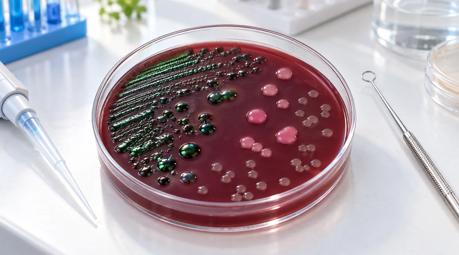
A student pulls an EMB plate from the incubator and stops. Several colonies are almost black with a green metallic shine. The student has two theories. Either the culture is contaminated, or the bacteria made green pigment. Both theories are wrong, and both come from the same missing concept.
An eosin methylene blue agar plate, usually shortened to EMB agar or emb plate, is a solid bacterial culture medium that contains the dyes eosin Y and methylene blue plus nutrients and agar. It is not a bottle of stain diluted into agar. It is a formulated system in which the same two dyes both suppress unwanted organisms and report sugar fermentation by changing colony color. Once that dual role is clear, the dark colonies and the occasional green sheen stop looking like accidents.
The short answer
EMB agar is selective against many Gram positive bacteria and differential for vigorous lactose fermentation. Eosin Y and methylene blue inhibit Gram positives, while acid from lactose fermentation drives precipitation of an eosin and methylene blue dye complex inside colonies. Strong acid producers such as many Escherichia coli strains turn dark blue black and may show a metallic green sheen in reflected light. Weaker fermenters turn pink to purple. Nonfermenters stay colorless or translucent. EMB is a complex, undefined medium because it contains peptone, and peptone supplies most of the nitrogen. Standard EMB does not contain sorbitol. Sorbitol belongs to a different screening task, mainly sorbitol MacConkey agar for E. coli O157:H7.
EMB is a medium, not a stain
The name causes the first confusion. Eosin methylene blue sounds like a staining solution, and methylene blue is indeed a cationic dye used for microscopy and for simple bacterial staining. But an EMB plate contains far more than dissolved dye. The working system includes a nitrogen and nutrient base, a defined fermentation sugar, a phosphate buffer, a solidifying agent, and the two dyes at low, carefully balanced concentrations.
That balance matters. The same methylene blue that colors cells on a slide behaves differently when paired with eosin Y in agar at near neutral pH and then exposed to local acid around a colony. For the broader dye family and why catalog names mislead, see methyl blue versus methylene blue. For how EMB compares with the other major enteric screen, see EMB agar versus MacConkey agar.
Two functions in one pair of dyes
Microbiology teaching separates two medium functions that EMB happens to combine. A selective medium suppresses some organisms while permitting a target group to grow. A differential medium lets organisms that do grow display visibly different reactions, usually through metabolism of one substrate. Standard teaching texts such as the OpenStax microbiology chapter on culture media define both terms, and EMB is a standard example of a medium that is both at once.
On EMB, selection comes from the dyes. Eosin Y and methylene blue together inhibit many Gram positive organisms, so enteric Gram negatives dominate the plate. The ASM eosin methylene blue agar protocol describes the medium as both selective and differential and emphasizes methylene blue as the stronger Gram positive inhibitor. Inhibition is partial rather than absolute, so occasional small Gram positive colonies can break through.
Differentiation comes from fermentation plus the same dyes. Lactose fermenters release acids around the colony. As local pH falls, eosin and methylene blue associate into a dye complex that deposits in the colony. Weak or moderate acid gives pink, brown, or purple growth. Strong acid gives dark blue black growth and, when deposition is heavy enough, the metallic green sheen by reflected light. The green is an optical effect of deposited dye, not a pigment secreted by E. coli. A useful chain to memorize is sugar fermentation, then acid, then lower pH, then dye complex deposition, then dark colony, then possible sheen.
Do not invert the two roles. EMB does not select lactose fermenters. Lactose use is the differential property. Dye inhibition is the selective property.
Is EMB agar defined or undefined
Students often ask whether emb agar is a defined or undefined medium. The answer is undefined, which is also called complex.
A chemically defined medium lists every chemical species at an exact concentration. A complex or undefined medium contains a biological digest or extract whose molecular makeup varies by batch. EMB contains peptone at 10 g per liter, and peptone is a protein hydrolysate with a mixture of peptides, amino acids, minerals, and growth factors. Naming the amount of peptone does not make the medium defined, because the composition of peptone itself is not fully specified.
This classification is independent of selective and differential behavior. EMB is complex, selective, differential, and solid at the same time. Those four labels answer four different questions: what is known about composition, who is allowed to grow, whether growers look different, and what physical state the medium has.
Cited formulation: Levine EMB
Recipes vary, so record which EMB the bench stocks. Two formulations dominate teaching and food microbiology. The older Holt-Harris-Teague style contains both lactose and sucrose. Levine EMB contains lactose without sucrose, which makes its differential reaction specific to lactose. The FDA BAM method for Levine EMB agar lists the Levine composition used in food testing.
| Ingredient per liter | Levine L-EMB, FDA BAM M80 |
|---|---|
| Peptone | 10 g |
| Lactose | 10 g |
| Dipotassium phosphate | 2 g |
| Eosin Y | 0.4 g |
| Methylene blue | 0.065 g |
| Agar | 15 g |
| Final pH | 7.1 plus or minus 0.2 |
The ASM student protocol prints a combined Difco style recipe with 10 g peptone, 5 g lactose, 5 g sucrose, 2 g dipotassium phosphate, 13.5 g agar, 0.4 g eosin Y, and 0.065 g methylene blue at pH 7.2, and ASM notes that it merges Holt-Harris-Teague sugars with Levine peptone and phosphate. That detail has practical force. On two sugar EMB, a dark colony may reflect sucrose use, lactose use, or both. On Levine EMB, a dark colony points to lactose. Proteus behavior illustrates the point: ASM warns that Proteus can show a sheen on sucrose containing EMB when spreading is suppressed, while on Levine EMB it usually stays colorless. Check the bottle or insert before interpreting.
Which ingredients supply nitrogen and carbon
The common exam question asks which ingredients in emb agar supply nitrogen. The main answer is peptone. Peptone provides peptides and amino acids, hence organic nitrogen, along with some carbon, minerals, and growth factors. It is the nutritional foundation of the plate.
The deliberately added carbohydrate is the carbon and energy source for the differential reaction: lactose in Levine EMB, or lactose plus sucrose in two sugar EMB. Dipotassium phosphate supplies phosphate and buffers pH so that only meaningful fermentation drops local pH enough to precipitate dye. Agar contributes no nutrition for this purpose. It only solidifies the plate. Eosin Y and methylene blue are the selective and reporting system.
Reading the plate: annotated chart
Read EMB under reflected or oblique light, because the sheen is easiest to see that way. Compare every unknown against known controls on the same batch. The chart below is an illustrative reconstruction for learning the pattern, not a photograph of live cultures.
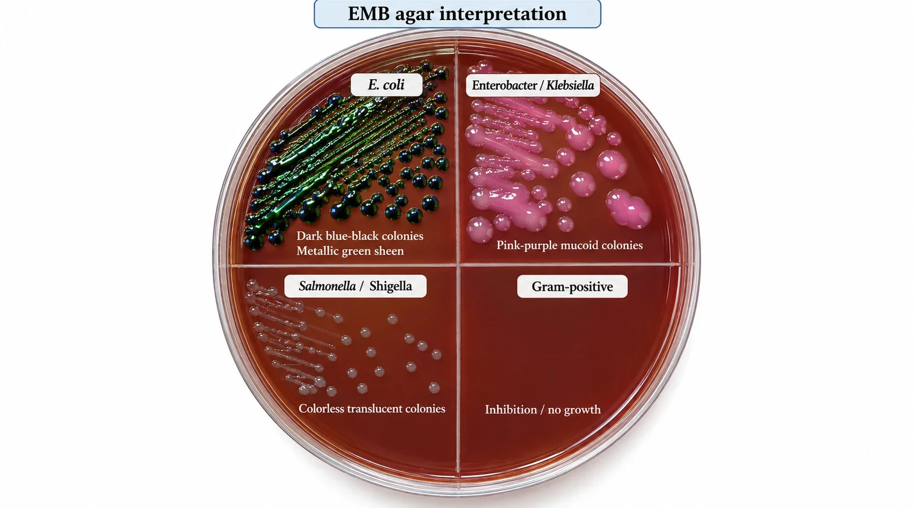
Use this reading logic:
- Dark blue black colonies, classically with a metallic green sheen: vigorous lactose fermentation, presumptive E. coli pattern. Confirm, because absence of sheen does not exclude E. coli and other strong acid producers can sheen on some formulations.
- Pink, brown, purple, often mucoid colonies without the classic sheen: slower or weaker fermentation, the usual Enterobacter and Klebsiella pattern.
- Colorless, translucent, pale, or amber colonies: nonfermenter pattern, the usual Salmonella, Shigella, and Proteus appearance on Levine EMB.
- No growth or only pinpoint colonies where Gram positives were streaked: expected selective suppression. Breakthrough growth happens and must not be read as a differential result.
Color is a screening phenotype. It directs Gram stain, oxidase, biochemical panels, or instrument identification. It never names a species alone. A direct side by side with the other enteric screen helps here, which is why a separate comparison covers EMB versus MacConkey selection and reading in detail.
Preparation overview
For dehydrated commercial EMB, follow the manufacturer insert for the exact product, since dye content and sterilization directions differ. For the ASM style formulation, the ASM eosin methylene blue agar protocol directs dissolving the ingredients, adjusting pH, boiling to dissolve agar, autoclaving 15 minutes at 121 degrees Celsius, cooling toward 60 degrees, swirling precipitate back into suspension, and pouring. The FDA Levine method specifies sterilization at no more than 121 degrees and the same final pH window. Some precipitate in EMB is normal and is resuspended by gentle swirling rather than discarded as contamination.
Incubate aerobically, typically 18 to 24 hours near 35 to 37 degrees, and read at the laboratory specified hour. Quality control commonly uses E. coli for the dark sheen reaction, a Salmonella strain and Proteus mirabilis for colorless growth, Enterobacter or Klebsiella aerogenes for mucoid fermentation, and Staphylococcus aureus plus Enterococcus faecalis to check Gram positive suppression. Match strain numbers and acceptance criteria to the insert.
What sorbitol EMB questions really ask
Another frequent exam prompt states that emb agar containing sorbitol is used to detect some pathogen, and asks which one. The premise needs correction before answering.
Standard EMB does not contain sorbitol. The established sorbitol screen for E. coli O157:H7 is sorbitol MacConkey agar, especially cefixime tellurite sorbitol MacConkey. The logic is that most ordinary E. coli ferment sorbitol rapidly and turn pink red, while typical O157:H7 strains ferment sorbitol slowly or not at all and stay colorless or gray, as described in the FDA BAM method for sorbitol MacConkey agar. There is no equivalent standardized sorbitol EMB in ASM or FDA bacteriological methods. In FDA diarrheagenic E. coli workflows, Levine EMB appears alongside chromogenic and other confirmatory media, but EMB alone cannot separate pathogenic STEC from ordinary E. coli. So when a question mentions sorbitol plus E. coli O157:H7, the intended medium is sorbitol MacConkey, not EMB. General molecular context for methylene blue itself sits in how methylene blue works.
Pitfalls that repeat every term
First, overreading the sheen. FDA describes typical E. coli on Levine EMB as dark centered with or without a sheen. Treat sheen as a strong presumptive clue, never as proof.
Second, ignoring formulation. A sucrose containing plate broadens the meaning of every dark colony. A Levine plate narrows it to lactose. Write the formulation in the notebook with the result.
Third, reading mixed growth as one organism. In mixtures, one organism can consume the acids made by another, which can erase the expected sheen. ASM documents such an E. coli and Salmonella mixture effect. Pick isolated colonies and work each morphotype separately.
Fourth, reading too late. Prolonged incubation lets weak fermenters and breakthrough Gram positives accumulate color and blurs the fermenter versus nonfermenter split. Record incubation time with the colony description.
Fifth, treating suppression as sterility. EMB reduces Gram positives. It does not guarantee their absence. A small colony in the Gram positive control streak is a product property to record, not necessarily a failed student.
Remember one sentence at the bench. Dyes select by suppressing Gram positives, acid from lactose speaks through dye deposition, and formulation decides which sugar the color is allowed to report.