
Article
Methylene blue electrochemistry: electrodes, redox probes, and sensors
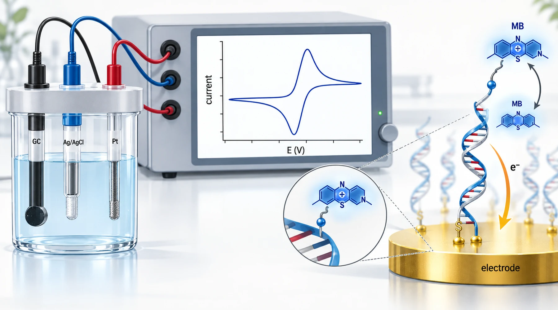
A graduate student runs a cyclic voltammogram of methylene blue on a glassy carbon electrode, sees a clean pair of peaks, and concludes the sensor is finished. A week later the peaks have drifted, shrunk, or split, and nothing in the protocol explains why. The dye did not change. The concept behind the measurement was incomplete.
Methylene blue earns its place in electrochemistry for three reasons. It exchanges electrons reversibly at mild potentials, it sticks to surfaces and polymerizes into films, and it binds nucleic acids. Each is a separate concept, and each fails in its own way. This review separates them: the couple, the readout, the modified electrode, the mediator role, and the sensor architectures built on top.
One couple, two forms, one proton
The core reaction at neutral pH is usually written as one oxidized cation taking up two electrons and one proton to give the reduced, colorless leuco form:
MB+ + H+ + 2e- gives LMB
The methylene blue redox potential at pH 7 sits near +11 mV against the standard hydrogen electrode, as reported in a recent mechanistic study of the dye in Chemistry Europe. Because one proton accompanies two electrons, the formal potential shifts about 30 mV negative for each pH unit increase. That slope is the first diagnostic worth memorizing. If your peak moves roughly 30 mV per pH unit, you are watching the expected couple. If it moves 60 mV per unit, the proton count has changed. If it barely moves at all, the signal may come from a surface film with its own acid base chemistry rather than dissolved dye.
Reference electrodes are the second habit. Potentials quoted against Ag/AgCl, SCE, and NHE differ by tens to hundreds of millivolts, and the filling solution shifts the numbers further. Every potential in this article names its reference. Any paper that does not is not reusable. The structural difference between the two forms, flat cation versus flexible neutral molecule, is itself part of the story, and the companion page on the redox pair treats it in detail.
What a voltammogram of methylene blue actually shows
On a clean bare glassy carbon electrode in roughly neutral phosphate buffer, dissolved methylene blue typically gives an anodic peak near 0.15 to 0.18 V negative of Ag/AgCl and a cathodic peak near 0.20 to 0.25 V negative of the same reference. The exact positions move with scan rate, surface history, and the reference filling solution, and a recent open access measurement on bare glassy carbon places the cathodic peak near 0.18 V negative at pH 7.4 under its own conditions.
The pair is best described as quasi-reversible. An ideal two electron reversible couple would show peaks about 30 mV apart. Methylene blue waves are generally wider and asymmetric, and adsorption complicates the picture. That complication is diagnostic, not noise. A peak current that grows in direct proportion to scan rate points to adsorbed dye. A peak current that grows with the square root of scan rate points to dye diffusing in from solution. Many real traces mix both, which is why the same solution can give different peak heights on a freshly polished electrode and a used one.
Dissolved oxygen contributes its own reduction current in the same window, so quantitative work is usually done after deoxygenation or with oxygen accounted for. And methylene blue fouls: adsorbed dye changes the surface with every cycle, so peak drift across repeated scans is evidence about the electrode, not about the analyte.
From dissolved dye to a modified electrode
A methylene blue modified electrode biosensor starts from a simple observation. The dye adsorbs strongly to carbon, so dipping an electrode in dye solution already modifies it. Simple adsorption is also the least stable construction, because the dye leaches back into solution. Everything else in this section is a response to leaching.
The most established answer is electropolymerized poly(methylene blue). Cycling the electrode oxidatively in dye solution deposits a redox active film that stays put far better than adsorbed monomer. The standard Karyakin type protocol uses about 1 mM dye in 0.02 M borate buffer near pH 9.1 with 0.1 M potassium chloride, cycling from about 0.40 V negative to 1.20 V positive for around 30 cycles, as documented in a Durham thesis record of the method. The alkaline buffer matters: film growth proceeds through the oxidized forms generated at positive potentials, and the window must reach them.
Other constructions solve different problems. Carbon paste and screen printed electrodes trade precision for disposability. Composites with nanotubes or graphene raise the active area and sharpen peaks. Gold electrodes with thiol anchored probes serve the nucleic acid sensors below. Entrapment under Nafion films slows leaching at the cost of slower response. The design question is always the same: which failure mode are you buying off, and what did you pay for it.
The mediator concept: a relay, not a reagent
As a methylene blue cyclic voltammetry redox mediator, dissolved dye acts as an electron relay between a reluctant analyte and the electrode. The oxidized dye accepts electrons from the sample chemistry, the electrode reoxidizes the reduced dye, and the measured current reports the turnover. Typical freely diffusing concentrations are tens of micromolar, roughly 10 to 60, rather than any fixed standard.
Three mediated chemistries recur. The dye mediates NADH oxidation, including enzyme coupled turnover, and poly(methylene blue) films electrocatalyze the same reaction at the surface. With oxidases it can serve as an artificial electron acceptor. With peroxidase systems, reduced dye shuttles electrons to peroxide reducing intermediates, which permits hydrogen peroxide detection at low applied potentials where interferences are fewer. A 2025 Chemical Science study of mediated NADH oxidation illustrates the mechanism in detail. At high dye loading the mediator background and oxygen chemistry can exceed the analytical signal, so mediator concentration is an optimization variable, not a reagent excess.
Nucleic acid sensors: where binding becomes current
Methylene blue binds DNA through a mix of intercalation, electrostatic attraction, and guanine specific interaction, so calling it simply an intercalator understates the chemistry. Sensor designs convert that binding into current in two distinct architectures, and confusing them is the most common reading error in this literature.
In label free designs, free dye in solution associates with surface bound probe DNA, and hybridization changes the signal. A common signal off format: single stranded probe accumulates dye and gives a large peak, while hybridization to double stranded DNA changes the dye environment and the signal drops. These formats routinely reach nanomolar detection limits. One example assembled complementary DNA on single walled carbon nanotubes on gold and reported linearity from 100 to 1000 nM with a detection limit near 7 nM in Electroanalysis.
In labeled designs, the dye is covalently attached to one end of the probe, the other end is thiolated to gold, and square wave voltammetry tracks the attached reporter. Folding based electrochemical aptamer sensors extend this idea: target binding rearranges the aptamer and moves the reporter relative to the electrode. An ochratoxin A sensor using a thiol and dye dual labeled aptamer on gold reported a range of 0.1 to 1000 pg per mL with a detection limit near 0.095 pg per mL, published in Analytica Chimica Acta.
The distinction matters for troubleshooting. In label free formats the dye is a solution component with its own concentration and pH dependence. In labeled formats the dye is part of the probe, and signal changes report distance and dynamics, not dye uptake.
Enzymatic and composite examples
Poly(methylene blue) films also anchor enzymatic sensors. One ammonium sensor combined a screen printed electrode with gold nanoparticles and a dye film, then coated glutamate dehydrogenase chemistry on top. The film oxidizes NADH while the enzyme consumes it in proportion to ammonium: 0 to 300 micromolar with a detection limit near 0.65 micromolar, reported in Biosensors. The full landscape is covered in a 2025 review of the dye in electrochemical sensing in Analytica Chimica Acta.
Conditions that travel with every result
The sibling reference on quantitative assay practice covers calibration discipline generally. For electrochemical results specifically, these entries belong in every lab record:
| Entry | Why it matters | Typical value here |
|---|---|---|
| Reference electrode and filling solution | Defines the potential axis | Ag/AgCl (3 M KCl), or convert explicitly |
| Buffer, pH, ionic strength | Peak position moves ~30 mV per pH unit | Phosphate buffer, pH 7.0 to 7.4 |
| Dye state: dissolved, adsorbed, or polymerized | Each has different stability and proton chemistry | Tens of micromolar dissolved, or Karyakin film |
| Scan rate and technique | Distinguishes adsorption from diffusion control | CV at 50 mV per s, SWV for labeled probes |
| Atmosphere | Oxygen reduces in the same window | Deoxygenated or oxygen corrected |
| Surface history | Fouling shifts every subsequent scan | Polish, cycle count, and storage stated |
A reporter is not a therapy
The final distinction is the one this page exists to enforce. A falling square wave peak on hybridization proves that a redox label moved or that dye binding changed. It proves nothing about what methylene blue does in a patient. Measurement chemistry and biological activity share a molecule and share nothing else: different concentrations, different matrices, different counter electrodes replaced by membranes and enzymes. Keep therapeutic questions on the biological activity pages, and keep sensor claims tied to the electrode, buffer, and technique that produced them. A redox label is a reporter. Reporters describe the assay, not the body.