
Article
Methylene blue and Bartonella: laboratory findings and clinical-evidence limits
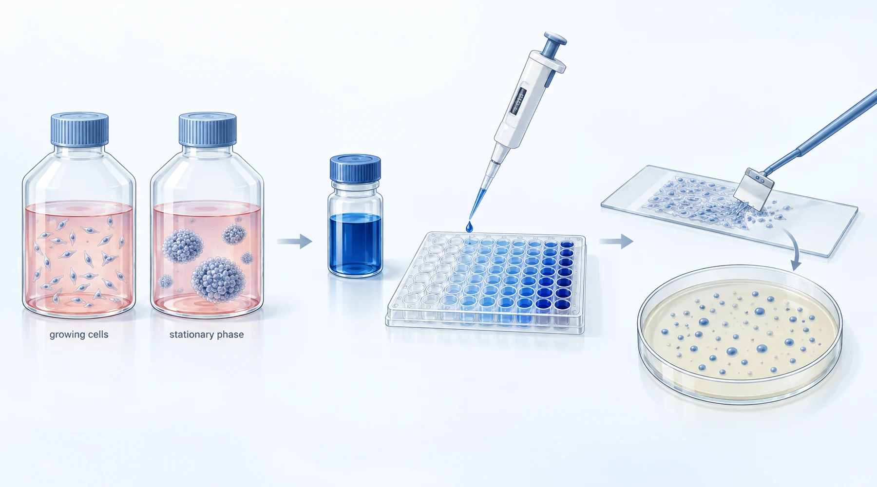
A culture dish and a patient share a word, infection, but not a concept. In the dish, infection is a count of colonies on agar. In the patient, it is bacteria inside cells and vessels, drugs moving through blood and tissue, and an immune system doing part of the work. Confuse the two and a striking laboratory number starts to look like a treatment. Keep them separate and the same number becomes what it is: a reason to do the next experiment.
That separation is the whole job of this review of methylene blue and Bartonella. Two laboratory studies from one research group tested methylene blue against Bartonella henselae in non-growing states. Both reported strong activity in glass and plastic. Neither tested a person.
Why researchers test the stationary phase
Most antibiotic testing uses growing cells. That model misses a harder case: cells that have stopped dividing and tolerate drugs that kill their growing siblings.
Bartonella can persist in the body, including inside erythrocytes and endothelial cells. So the group asked which approved drugs can kill B. henselae after it has entered stationary phase. A low MIC against growing cells did not predict stationary-phase killing. Rifampin had a very low MIC yet killed stationary cells poorly, while compounds with higher MIC values killed them well. Readers who want a general method for this kind of paper will find it in the guide to reading methylene blue research.
First screen: Li and colleagues, 2019
The 2019 stationary-phase screen in Antibiotics used a single strain, B. henselae JK53, grown in Schneider medium with 10 percent fetal bovine serum at 37 C with 5 percent CO2. One-day cultures were mostly dispersed dividing cells. Five-day cultures, at around 10 to the 8th colony forming units per mL, were taken as stationary phase and contained visible aggregates.
The assay was SYBR Green I with propidium iodide, calibrated against mixtures of live and isopropanol-killed cells. Because methylene blue is strongly colored, hits were confirmed by microscopy and then by colony counts on blood agar, the stricter endpoint.
From 1,581 approved drugs at 50 micromolar, methylene blue left 16 percent viability by plate reader and 25 percent by microscopy. Daptomycin scored 21 percent, several azoles scored 22 to 27 percent, gentamicin 32 percent, erythromycin 66 percent, ciprofloxacin 74 percent. Pyrvinium pamoate looked stronger on the reader but could not be scored by microscopy because of its own fluorescence.
Against growing B. henselae the methylene blue MIC was 0.08 to 0.16 micrograms per mL. Against five-day stationary cells at 5 micrograms per mL, the colony count fell to no detectable colonies after one day of exposure, matching daptomycin and pyrvinium pamoate at their tested concentrations. A separate time-kill run used 10 micrograms per mL. Those are laboratory exposure levels, not doses, and the paper states them as such. A cited human peak plasma value near 3.9 micrograms per mL appears in the paper only as background context. The overview of methylene blue pharmacokinetics explains why a plasma number cannot be read as a tissue number.
Two limits are explicit in the paper. There was no biofilm model and no methylene blue combination testing. The five-day aggregates were clusters in stationary liquid culture, not a validated biofilm experiment. The authors called for animal work, combination work, biofilm work, and testing in other Bartonella species.
Combinations and biofilm-derived cells: Zheng and colleagues, 2020
The 2020 follow-up in BMC Microbiology kept the same strain and medium but moved to six-day stationary cultures and added two things the first paper lacked: two-drug combinations and a biofilm-derived cell model.
For single drugs at a uniform 5 micrograms per mL for 24 hours, methylene blue left 3.2 times 10 to the 4th colony forming units per mL against a control near 2.8 times 10 to the 10th. Ciprofloxacin, gentamicin, and nitrofurantoin left fewer colonies at the same point. That differs from the 2019 result, where the same nominal level gave no detectable colonies after one day. Culture age, density, and handling differed, so killing was reproducible in direction but not identical in magnitude. This run-to-run shift is one reason standard safety and interaction checks treat laboratory potency as separate from clinical effect.
The combination screen is the most quoted part. Every component was fixed at 5 micrograms per mL. Both azithromycin plus methylene blue and rifampin plus methylene blue gave no detectable stationary-phase colonies at 24 hours, as did both ciprofloxacin pairs, and only those four among 25 tested pairs. No formal synergy analysis was run, so the correct term is active combination under these conditions, not proven synergy.
The biofilm experiment tested cells recovered from a young disrupted biofilm, not penetration into an intact mature one. Cultures were grown five days, diluted into well plates for five more days, confirmed by crystal violet staining, then scraped before drug exposure. At 5 micrograms per mL, methylene blue alone fell to 2.3 times 10 to the 2nd by day 6, among the best single agents, but not zero. Both methylene blue combinations reached zero by day 6, as did both ciprofloxacin combinations.
The authors flagged the same limits a careful reader would: young scraped biofilm rather than old intact biofilm, one convenient concentration for all drugs instead of each drug at its own realistic peak, only selected pairs centered on azithromycin or rifampin, one strain, no animals, no patients. Later three-drug work was proposed, not reported.
Study-design comparison
| Design point | Li 2019 | Zheng 2020 | What counts as clinical evidence |
|---|---|---|---|
| Organism | B. henselae JK53 only | B. henselae JK53 only | Patients with confirmed bartonellosis, defined endpoints |
| Growing-cell MIC for methylene blue | 0.08 to 0.16 micrograms per mL | Same, 0.08 to 0.16 | Not applicable |
| Stationary model | 5-day culture, single drugs | 6-day culture, single drugs plus 25 pairs | Tissue and intracellular activity, pharmacokinetics |
| Biofilm model | None, aggregates are not biofilm | Scraped 5-day biofilm-derived cells | Intact biofilm or in vivo persistence data |
| Key methylene blue result | 5 micrograms per mL, no colonies at day 1 | Same level, 3.2 times 10 to the 4th at 24 hours; pairs with azithromycin or rifampin at zero | Response rate, relapse rate, adverse events |
| Formal synergy test | Not done | Not done | Dose finding and controlled comparison |
The table is the point of this article in compact form. Everything left of the last column is culture work. Nothing in either paper occupies the last column.
Bartonella is not Borrelia
Both papers mention Borrelia burgdorferi, the Lyme disease spirochete, because the dye assay came from earlier Borrelia stationary-phase work in the same laboratory, where methylene blue and daptomycin had also scored as hits. That history is useful context and a common source of error.
No experiment in either Bartonella paper tests Borrelia side by side with Bartonella. Organisms differ, media differ, concentrations differ, and the hypothesized membrane effect of methylene blue in Bartonella was stated as speculation needing more study. Results should not be carried across species, and neither paper supports broad claims about parasites. The comparison of Borrelia and Bartonella persisters keeps the two literatures apart for this reason.
Why cultures do not convert into a protocol
Search interest in a methylene blue bartonella protocol is understandable. These papers are why that phrase should be treated as a warning flag rather than a shopping list.
First, 5 micrograms per mL in a well is not a prescription. It has no absorption, distribution, protein binding, intracellular penetration, or tolerability attached. The authors chose it as a rough average of antibiotic peaks and said future work should use each drug at its own realistic level.
Second, zero colonies means no colonies under those plating conditions, not sterile tissue. Persisters, intracellular bacteria, and sampling limits all sit outside the agar plate.
Third, combinations that sterilize a well can fail or harm in a body. Azithromycin plus methylene blue and rifampin plus methylene blue are laboratory pairs at laboratory exposures. Without animal pharmacokinetics and controlled human data there is no dose, no duration, and no benefit-to-risk ratio. Anyone weighing options should discuss them with a clinician and review when methylene blue use needs medical supervision before drawing any conclusion from dish data.
The narrow conclusion these papers do support is this: methylene blue has reproducible activity against non-growing B. henselae JK53 in vitro, including in two-drug laboratory combinations, with the strongest combination and biofilm-derived results coming from the 2020 study. Everything else, including whether that activity matters for human bartonellosis, remains untested.