
Article
EMB agar versus MacConkey agar: selecting and interpreting media
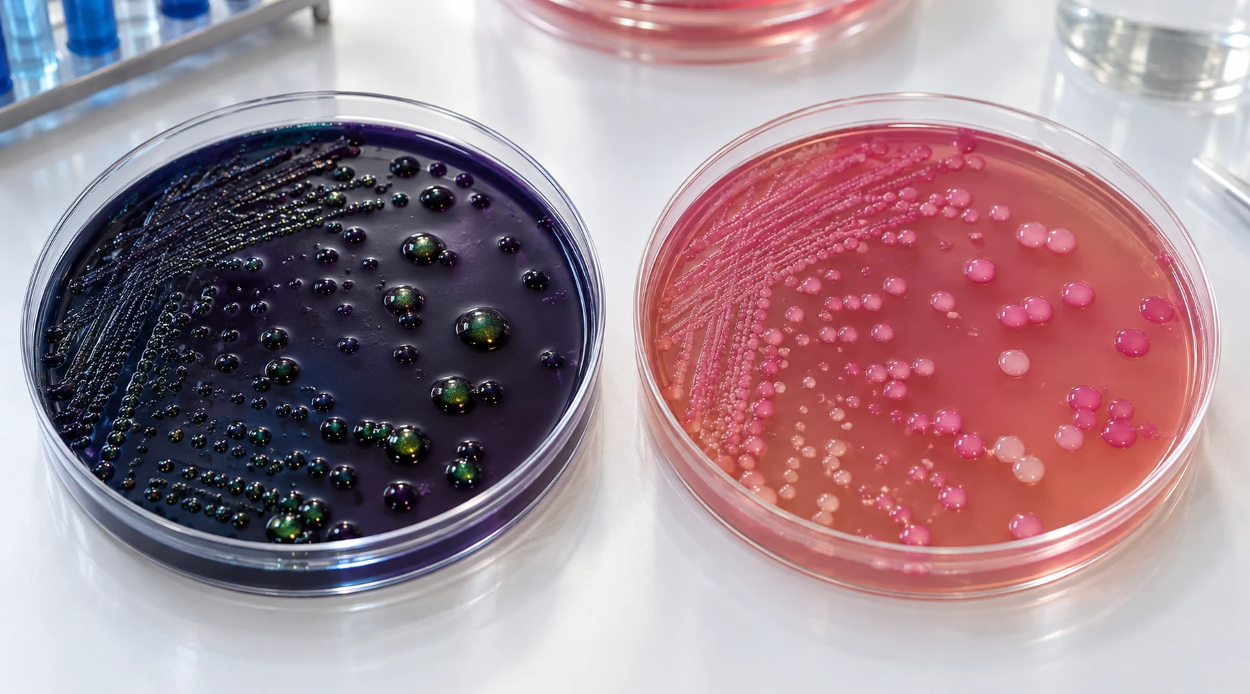
A student streaks the same water sample on two plates. On one, dark colonies with a green metallic shine appear. On the other, bright pink colonies with a hazy halo appear. Both plates were inoculated from the same tube, incubated side by side, and read at the same hour. The student asks which plate is right.
Both plates are right. They answer different questions. Confusion comes from treating the two media as interchangeable screens for gut bacteria. They share a purpose, which is to select Gram negative enteric organisms and separate lactose fermenters from nonfermenters, but they implement that purpose with different chemistry and different visual logic. Once you see the two underlying concepts, plate selection and colony reading become routine.
The short answer
EMB agar uses two dyes, eosin Y and methylene blue, that do double duty: they suppress Gram positive growth and they precipitate around strong acid producers to create dark colonies, classically with a metallic green sheen for Escherichia coli. MacConkey agar separates the jobs: bile salts plus crystal violet suppress Gram positives, lactose feeds fermentation, and neutral red reports acid as pink or red colonies. If the task is a vivid presumptive demonstration of E. coli, EMB earns its place. If the task is a clean pink versus colorless sort of lactose fermenters from nonfermenters, especially in stool or urine workflows, MacConkey is usually the simpler read. Neither plate identifies an organism by color alone.
EMB: dyes that select and report at once
The EMB concept packs selection and indication into the same molecules. The ASM eosin methylene blue agar protocol describes EMB as both selective and differential, with the dye pair inhibiting many Gram positive organisms while lactose fermentation drives dye uptake and precipitation. A standard Levine formulation carries peptone, lactose, phosphate, eosin Y, methylene blue, and agar, as listed in the FDA BAM method for Levine EMB agar. Note that older EMB recipes sometimes added sucrose alongside lactose, while Levine EMB contains lactose without sucrose, so record which formulation your laboratory stocks.
Reading EMB means reading dye behavior. Vigorous lactose fermentation produces enough acid to drive heavy dye precipitation, giving E. coli its dark blue black centers and, in the classic case, a green metallic sheen in reflected light. Weaker fermenters such as Enterobacter and Klebsiella typically grow as pink to purple mucoid colonies without that sheen. Nonfermenters such as Salmonella, Shigella, and Proteus generally grow as colorless or translucent colonies because they do not produce the acid burst that precipitates the dyes. Gram positives are usually suppressed, though suppression is a matter of degree and varies by strain and product. Full dye preparation chemistry, including why methylene blue behaves as a cationic stain in the first place, belongs to a separate discussion of dye formulations, and the general microscopy context sits in a staining methods atlas. This article stays on plate selection and reading.
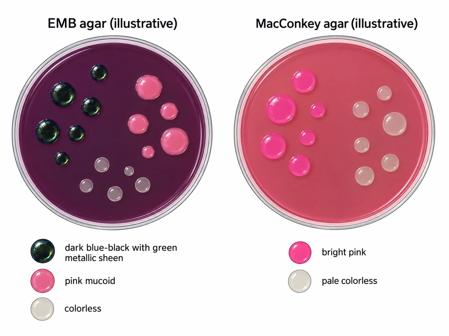
The figure above is illustrative. It shows the visual logic to expect: dark sheen colonies plus pink mucoid types plus pale types on EMB, against pink versus pale types on MacConkey. Use it to learn the pattern, then confirm every call against photographed controls and the product insert for the exact medium on your bench.
MacConkey: separated jobs, simpler visual rule
The MacConkey concept assigns each job to a separate ingredient. The ASM MacConkey agar protocol presents the same selective and differential framing, and the FDA BAM method for MacConkey agar lists the working parts: peptones, lactose, bile salts, salt, neutral red, crystal violet, and agar. Bile salts plus crystal violet select against Gram positives. Lactose supplies the fermentation substrate. Neutral red acts as the pH reporter, turning pink to red where acid forms, sometimes with precipitated bile around strong fermenters.
The reading rule is deliberately plain: pink means lactose fermenter, colorless or pale means nonfermenter. E. coli appears pink to red, often with a bile precipitate halo. Klebsiella and Enterobacter appear pink and frequently mucoid because of their capsules. Salmonella, Shigella, Proteus, and many other Gram negative rods appear colorless. A side benefit matters in teaching labs: bile salts generally reduce Proteus swarming, so isolated colonies are easier to obtain on MacConkey than on nonselective media, though suppression is not absolute and salt free alternatives exist where swarming control is the priority.
The molecular reason the dyes behave this way traces to charge and structure. Methylene blue is a cationic phenothiazine dye whose structure and properties explain its affinity for acidic bacterial surfaces, which is the same affinity at work when it precipitates in acid zones on EMB.
Comparison at a glance
| Feature | EMB agar | MacConkey agar |
|---|---|---|
| Selective system | Eosin Y plus methylene blue | Bile salts plus crystal violet |
| Differential substrate | Lactose (Levine has no sucrose) | Lactose |
| Indicator | Dye precipitation itself (dark colonies, green sheen) | Neutral red (pink to red acid colonies) |
| Strong E. coli signal | Dark blue black colony, often metallic green sheen | Pink to red colony, often bile precipitate |
| Klebsiella and Enterobacter | Pink to purple, mucoid, usually no sheen | Pink to red, often mucoid |
| Salmonella, Shigella, Proteus | Usually colorless or translucent | Usually colorless or pale |
| Gram positives | Usually suppressed | Usually suppressed on crystal violet formulas |
| Best teaching payoff | Strong versus weak fermentation made visible | Simplest fermenter versus nonfermenter sort |
Read the table as a decision procedure. When the exercise needs students to see a gradient of fermentation strength with one dramatic endpoint, EMB shows it. When the exercise needs a fast binary sort before biochemical confirmation, MacConkey shows it.
Which medium for which sample
For water exercises aimed at presumptive fecal coliform or E. coli recognition, EMB is attractive because the sheen is visually distinctive. MacConkey also handles enteric Gram negatives and has historical water use, but neither plate substitutes for a regulatory water method, which may specify media such as modified mTEC or MI depending on the test. Check the method your laboratory is required to follow.
For stool style separations of fermenters from nonfermenters, MacConkey is generally the more useful of the two. Practical stool work for Salmonella or Shigella normally adds more selective enteric media such as XLD or Hektoen enteric rather than relying on MacConkey alone.
For urine style teaching that mimics clinical workflow, pair MacConkey with blood agar. MacConkey provides the early Gram negative sort while blood agar recovers organisms that MacConkey suppresses. This pairing is standard in clinical descriptions of urine culture setup.
For pure concept teaching, run both. EMB teaches that fermentation strength matters and that one molecule can serve two roles. MacConkey teaches modular design: selector, substrate, and reporter kept separate. Students who streak a known E. coli, a known Klebsiella aerogenes, and a known Salmonella side by side on both media rarely confuse the two logics again.
Reading pitfalls that repeat every term
The most common failure is identification by color alone. Pink on MacConkey can be E. coli, Klebsiella, Enterobacter, or another fermenter. Colorless can be Salmonella, Shigella, Proteus, Pseudomonas, or another nonfermenter. Color is a screening phenotype that directs confirmatory work such as Gram stain, oxidase, biochemical panels, or instrument identification, not a species name.
The second failure is overreading a missing sheen. Some genuine E. coli colonies, especially isolated ones, produce little or no metallic sheen on EMB. Absence of sheen does not exclude E. coli, and presence of sheen does not prove it. Treat the sheen as a presumptive clue.
The third failure is comparing plates of different ages. Delayed lactose fermenters can turn pink on MacConkey after prolonged incubation, which blurs the clean fermenter versus nonfermenter split. Read at the laboratory specified hour and record incubation time with the result.
The fourth failure is reading mixed growth as one organism. A pink colony next to a colorless colony records two phenotypes, not an intermediate. Pick well isolated colonies and work each morphotype separately. Mucoid texture adds a similar trap: both Klebsiella and Enterobacter can look mucoid, so capsule alone never settles the name. The organism once taught as Enterobacter aerogenes is now classified as Klebsiella aerogenes, which explains why older charts and newer labels seem to disagree.
Controls and a closing rule
Run photographed or fresh controls every session: E. coli for the strong fermenter reaction, Klebsiella aerogenes for mucoid fermentation without the classic sheen, a Salmonella strain for the colorless nonfermenter reaction, and Staphylococcus aureus to confirm Gram positive suppression on your exact product. Match strain numbers and acceptance criteria to the manufacturer insert, because selectivity shifts with bile, crystal violet, and dye content.
Remember one sentence at the bench. EMB uses dyes that both select and speak, which buys a dramatic E. coli signal at the cost of a subtler read. MacConkey separates selecting from speaking, which buys a plain pink versus pale sort at the cost of less fermentation detail. Choose the medium whose concept matches the question, confirm color with proper tests, and the two plates stop competing and start cooperating.