
Edemede
Nhọrọ ndị ọzọ maka methylene blue na mikroskopi: họrọ dịka ọrụ si dị
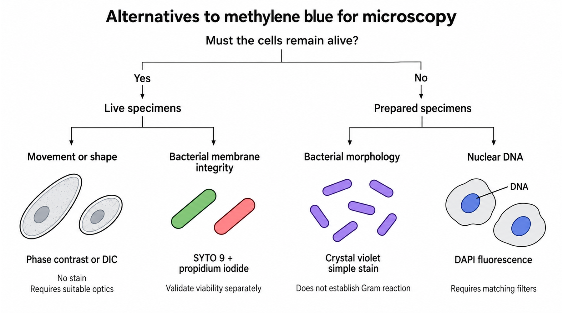
Maka bacterial smear dị mfe, crystal violet nwere ike dochie methylene blue mgbe ebumnuche bụ ịhụ ọdịdị sel. Maka sel dị ndụ, nhọrọ ka mma nwere ike ịbụ phase contrast na-enweghị stain ọ bụla. Nhọrọ ahụ dabere n'ihe ịchọrọ ileba anya.
Were ya na mmadụ abụọ na-enyocha otu bacterial culture. Otu chọrọ ịmata ọdịiche dị n'etiti sel yiri mkpara na sel gbara gburugburu. Nke ọzọ chọrọ ịma ma treatment egbuola bacteria. Stain nke na-enye onye mbụ onyonyo doro anya nwere ike ghara inye onye nke abụọ azịza bara uru.
Tupu ịhọrọ ihe ga-anọchi ya, mezue ahịrịokwu a: “Achọrọ m ileba anya na ___ n'ime ___, ma sel ndị ahụ ga-anọgide / agaghị achọ ịnọgide dị ndụ.” Ahịrịokwu ahụ na-akọwa ọrụ stain ahụ ga-arụ.
Malite na specimen na ajụjụ ahụ
Buru ụzọ kpebie ma sel ndị ahụ ga-anọgide dị ndụ, wee chọpụta feature ịchọrọ ileba anya:
- Sel ga-anọgide dị ndụ: maka mmegharị ma ọ bụ ọdịdị, tụlee unstained phase contrast ma ọ bụ DIC; maka nuclear DNA, tụlee Hoechst 33342 na fluorescence; maka membrane integrity, họrọ assay a kwadoro dịka SYTO 9 na propidium iodide.
- Sel agaghị achọ ịnọgide dị ndụ: maka bacterial morphology, tụlee crystal-violet simple stain ma ọ bụ nigrosin negative stain; maka Gram reaction, jiri complete Gram-stain method; maka nuclear DNA n'ime fixed cells, tụlee DAPI fluorescence.
Jiri tebụl a tụnyere oke nke ngalaba ọ bụla. Ndị a bụ nhọrọ usoro, ọ bụghị ntuziaka ka e jiri otu karama dochie nke ọzọ n'ime protocol dị adị.
| Ihe ịchọrọ ileba anya | Specimen na akụrụngwa | Nhọrọ ọzọ enwere ike iji | Oke kachasị mkpa |
|---|---|---|---|
| Ọdịdị na nhazi bacteria | Prepared bacterial smear; brightfield | Crystal violet simple stain | Na-egosi morphology; ọ naghị egosi Gram reaction ma ọ bụ viability. |
| Akụkụ mpụta sel megide background gbara ọchịchịrị | Bacteriological preparation; brightfield | Nigrosin negative stain | Preparation na formulation dị mkpa; sel na-enweghị stain abụghị n'ezie sel dị ndụ. |
| Gram reaction | Prepared bacterial smear; brightfield | Complete Gram-stain method | Chọrọ usoro reagent niile na controls. |
| Mmegharị ma ọ bụ mgbanwe n'ime sel dị ndụ | Compatible wet preparation; phase contrast ma ọ bụ DIC | Enweghị stain | Chọrọ optics kwesịrị ekwesị; contrast anaghị achọpụta molecule. |
| Nuclear DNA | Fixed cells; fluorescence | DAPI | Chọrọ excitation na emission filters dakọtara. |
| Nuclear DNA n'ime sel dị ndụ | Live cells; fluorescence | Hoechst 33342 | Ịbanye n'ime sel anaghị egosi na imaging conditions adịghị emerụ ahụ. |
| Bacterial membrane integrity | Unfixed bacteria; fluorescence | SYTO 9 na propidium iodide | Membrane staining bụ indirect measure nke viability. |
Maka simple smear, jiri nlezianya dochie usoro ahụ
Crystal violet bụ nhọrọ bara uru mgbe ọrụ ahụ bụ ịhụ bacterial morphology n'otu agba. FDA's Bacteriological Analytical Manual depụtara crystal-violet formulations abụọ kwesịrị ekwesị maka simple staining. Jiri formulation edekọtara na usoro preparation ya; aha dye naanị anaghị akọwa working reagent.
Crystal violet na-apụtakwa na Gram staining, ma nke ahụ anaghị eme crystal-violet simple stain ka ọ bụrụ Gram stain. N'ime ASM Gram-stain protocol, differential result dabere na crystal violet, iodine, decolorization, na counterstaining na-arụ ọrụ n'usoro. Ịhapụ usoro ndị ahụ na-agbanwe ihe onyonyo ahụ nwere ike igosi. Lee simple staining megide Gram staining maka ọdịiche ahụ.
Nigrosin na-enye ụzọ ọzọ isi mee ka akụkụ mpụta pụta ìhè: ọ na-eme ka background gbaa ọchịchịrị ebe bacterial cells na-anọgide na-enwu karịa ya. E nwere nkọwa bara uru n'ime ntuziaka nigrosin nke Merck nke ndepụta nkịtị nke ihe nnọchi nwere ike ileghara anya. Working formulation ya akọwapụtara gụnyere formaldehyde. Ya mere, ị gaghị ekpebi na ọ dabara na live cells naanị site n'okwu “negative stain,” ọbụlagodi na sel ndị ahụ n'onwe ha yiri ka ha enweghị stain. Lelee formulation zuru ezu na specimen preparation.
Maka specimen dị ndụ, tụlee ịgbanwe optics
Ọ bụrụ na ịchọrọ ịhụ sel ka ha na-agagharị ma ọ bụ na-ekewa, itinye dye ọzọ nwere ike ime ka observation sie ike. Dịka Nikon kọwara na ntuziaka ya gbasara contrast n'ime transparent specimens, phase contrast na differential interference contrast, ma ọ bụ DIC, na-eme ka specimen pụta ìhè site na ọdịiche dị na optical properties ha. Ha nwere ike igosi sel dị ndụ na-enweghị stain, ma ọ bụrụ na microscope nwere components achọrọ.
Nhọrọ a na-echekwa preparation na-enweghị stain, ma ọ na-agbanwe ozi enwere ike inweta. Ị nwere ike soro boundary ma ọ bụ mmegharị na-amaghị molecule nke kpatara contrast ahụ. Ọ bụrụ na ajụjụ ahụ gbasara molecular localization, họrọ labeling method kwesịrị ekwesị kama.
Maka nuclear DNA, ntụnyere DAPI na Hoechst stains nke Invitrogen na-ekewa ojiji DAPI a na-ahụkarị na fixed cells na Hoechst 33342 nke na-abanye sel karịa, nke a na-ahọrọ maka live-cell labeling. Ha abụọ chọrọ fluorescence equipment. Ọ dịghị nke ọ bụla bụ direct replacement maka colored brightfield stain.
Were dye exposure na illumination dịka experimental conditions. Tụnyere stained na unstained preparations mgbe behavior nke sel bụ measurement gị.
Sel uhie abụghị sel nwụrụ anwụ ozugbo
A na-ejikọkarị SYTO 9 na propidium iodide iji nyochaa bacterial membrane integrity. BacLight manual na-ejikọta intact membranes na green staining, yana damaged membranes na red staining. Interpretation ahụ chọrọ validation maka specimen ahụ.
Ọmụmụ 2019 nke Rosenberg na ndị ọrụ ibe ya na-egosi nsogbu ahụ. N'ime 24-hour E. coli biofilms n'elu glass n'ime phosphate-buffered saline, ihe dịka 96% nke sel pụtara propidium-iodide-positive in situ. Ma ndị ode akwụkwọ tụrụ aro na 82% ka nwere ike cultivable mgbe harvesting gasịrị. Confocal images gosiri sel nwere green interiors n'okpuru red-stained material, nke na-egosi extracellular nucleic acids dịka ihe kpatara signal na-eduhie eduhie.
Percentages ndị ahụ na-akọwa measurements dị iche iche emere na preparations nke ọmụmụ ahụ, ọ bụghị universal correction factor. Ihe bara uru ị ga-amụta bụ ịlele ihe mepụtara signal ahụ. N'ime biofilm, nucleic-acid dye nwere ike ịchọpụta material dị n'èzí sel yana nke dị n'ime damaged cells. Maka viability conclusion, ndị nchọpụta na-akwado complementary measurements, dịka cultivation ma ọ bụ metabolic activity, ebe ha na-ekweta na nke ọ bụla nwere limitations.
Lelee ihe nhọrọ ọzọ ahụ nwere n'ezie
Alternative staining method ka nwere ike ịnwe methylene blue. Giemsa bụ otu ihe atụ: Merck kọwara product ya dịka azure-eosin-methylene-blue solution, nke e mere maka applications gụnyere blood na bone-marrow smears. Ọ nwere ike ịbụ usoro dị iche maka specimen dị iche, ma ọ naghị emezu requirement nke iwepụ methylene blue.
Ọ bụrụ na iwepụ compound bụ akụkụ nke experiment, lelee reagent identity na formulation tupu ịtụnyere images. Ntuziaka gbasara methyl blue, methylene blue, na dyes ndị nwere aha yiri ibe ha na-akọwa ọdịiche dị n'aha.
Nyochaa substitution tupu iji result ahụ
Maka usoro ọhụrụ, mee obere ntụnyere site na representative specimens na appropriate control. Debe microscope settings ka ha bụrụ comparable ebe o kwere mee. Dekọọ reagent formulation, specimen preparation, exposure, na imaging conditions ka e wee nwee ike nyocha ihe mere otu image ji dị mma karịa kama ịhọrọ ya naanị n'ihi ọdịdị ya.
Tulee ihe ịga nke ọma dabere na ajụjụ mbụ. Maka morphology, nyochaa ma cell boundaries na arrangement ka nwere ike ịghọta nke ọma. Maka nuclear labeling, nyochaa signal location na background. Maka viability, chọọ ihe akaebe karịrị attractive color separation. Ntuziaka sara mbara gbasara methylene blue staining na laboratory applications na-akọwa otú observation na preparation si kwekọọ.
Họrọ nhọrọ ọzọ nke na-echekwa measurement ịchọrọ. Agba bụ naanị otu property nke image a ga-enweta.